
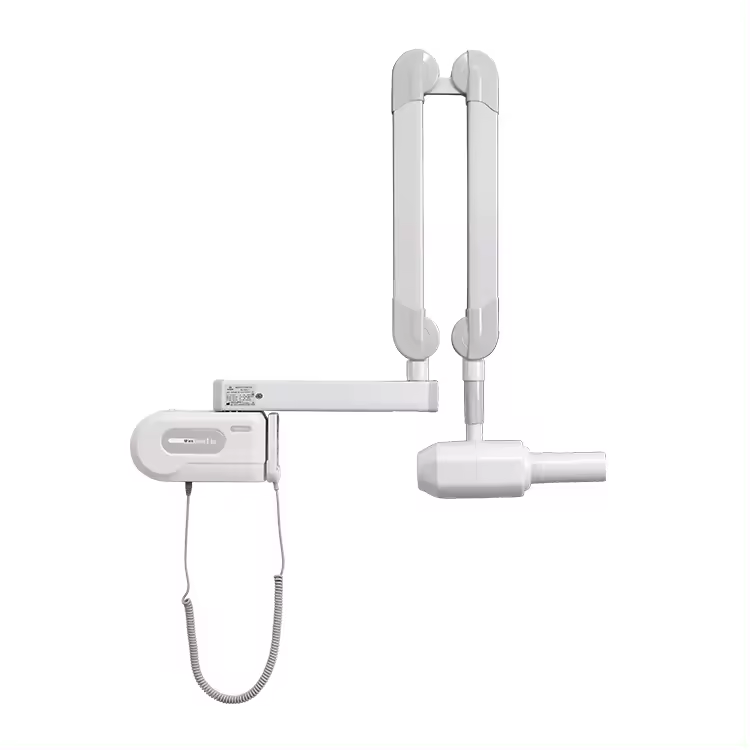


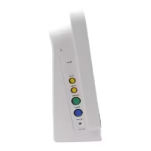
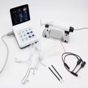
RVG Sensor
An RVG (RadioVisioGraphy) sensor is a digital imaging device used in dental radiography to capture high-resolution intraoral X-ray images quickly and efficiently, offering reduced radiation exposure and instant image display for enhanced diagnostic accuracy and patient comfort.
Description
RVG (RadioVisioGraphy) sensor
An RVG (RadioVisioGraphy) sensor is a digital imaging device used in dental radiography to capture intraoral X-ray images. Here is an overview:
RVG Sensor (RadioVisioGraphy Sensor)
- Definition: An RVG sensor is a type of digital sensor used to capture detailed images of teeth, bone, and surrounding oral structures directly onto a computer screen.
- Purpose: It replaces traditional X-ray film with a digital sensor, allowing for faster, clearer, and more efficient imaging.
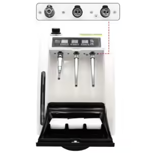
- Components:
- Sensor: A small, flat device that is placed inside the patient’s mouth to capture the X-ray image.
- X-ray Source: Emits X-rays that pass through the dental structures and are captured by the sensor.
- Computer and Software: The sensor is connected to a computer, and the image is processed and displayed using specialized software.
Key Features
- Digital Imaging: Converts X-ray energy into a digital image.
- High Resolution: Provides high-quality, detailed images for accurate diagnosis.
- Instant Image Display: Images are available immediately on the computer screen.
- Reduced Radiation Exposure: Requires less radiation compared to traditional film-based X-rays.
- Enhanced Image Processing: Software tools allow for image enhancement, zooming, and detailed analysis.
- Efficient Workflow: Streamlines the imaging process, saving time for both patients and dental practitioners.
- Storage and Sharing: Digital images can be easily stored, retrieved, and shared electronically.
Common Uses
- Cavity Detection: Identifying caries and other tooth decay issues.
- Root Canal Treatment: Assessing the internal structure of teeth.
- Implant Planning: Evaluating bone density and structure for dental implants.
- Periodontal Disease Assessment: Examining bone levels and detecting periodontal diseases.
- Orthodontics: Planning and monitoring orthodontic treatments.
Advantages
- Improved Diagnostics: High-quality images allow for more accurate and early diagnosis.
- Patient Comfort: The small size and shape of the sensor can be more comfortable for patients.
- Environmental Benefits: Reduces the need for chemical processing of X-ray films, making it more environmentally friendly.
- Data Management: Easy integration with electronic health records (EHR) and practice management software.
How It Works
- Placement: The sensor is placed inside the patient’s mouth, positioned near the area to be imaged.
- Exposure: The X-ray source emits X-rays that pass through the dental structures and are captured by the sensor.
- Image Capture: The sensor detects the X-rays and converts them into a digital signal.
- Image Processing: The digital signal is processed by the computer and displayed as a high-resolution image.
An RVG sensor is a valuable tool in modern dentistry, offering significant advantages over traditional film-based X-rays in terms of efficiency, image quality, and patient safety.


Reviews
There are no reviews yet.